Multimodal Biomedical Imaging Probes
Biomedical imaging is key for the diagnosis and monitoring treatment of disease states. Research into biomedical imaging of disease states, typically involves a series of in vitro, in vivo and/or ex vivo imaging experiments, where the spatial resolution involved ranges from the cellular level up to whole body imaging. However, no single imaging modality is capable of giving optimum imaging performance in all these scenarios.
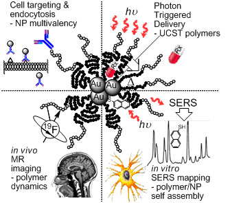
We are designing novel multimodal imaging probes with the aim to develop agents that are able to be used at all stages of biomedical imaging. As well as this we are actively developing ‘theranostics’, i.e. we are incorporating drug delivery mechanisms into the imaging agents. This is a multidisciplinary project that brings together researchers interested in fundamental polymer chemistry, nano- and biotechnology and pharmacy, as well as clinicians. The project involves multiple components, and these will be combined into a single device (see Figure).
- Design of multi-functional polymers
- This involves synthesis of amphiphilic polymers containing segments that exhibit an upper critical solution temperature (UCST). Development of new polymer systems that exhibit this behaviour will be utilized for photon-triggered drug delivery.
- Conjugation of drugs and targeting ligands
- Conjugation of common drugs (eg. Doxorubicin) and targeting ligands (eg. Folate) for cancer therapy will be investigated.
- Self-assembly around nanoparticles
- The self-assembly of a variety of polymer structures around gold nanoparticles will be investigated. Typically, the UCST responsive region of the amphiphilic polymers will be bound to gold nanoparticles – the construct will be designed to fall apart upon heating of the nanoparticles. The nanoparticle aggregates also act as a second imaging modality due to intense surface enhanced Raman scattering (SERS) that can be observed from the constructs. This modality is ideal for background free mapping of at the micron scale for in vitro and ex vivo experiments.
- Development of 19F MRI technology.
- 19F MRI is a new and emerging technology for contrast in magnetic resonance. We will incorporate recently developed MRI probes into the constructs for in vivo imaging and cell tracking.
- 19F MRI is a new and emerging technology for contrast in magnetic resonance. We will incorporate recently developed MRI probes into the constructs for in vivo imaging and cell tracking.
Collaborators
Peter Fredericks, Queensland University of Technology
Cameron Alexander, University of Nottingham
Source of Funding
ARC Discovery DP1094205
Point of contact: Idriss Blakey, Kris Thurecht